 Precision Machinery Technology Co., Ltd")


In different experimental scenarios, the objective and eyepiece lenses may need to be used in reverse. The Manual 3D Live Cell Imaging Microscope is capable of being flipped for use, facilitating the switch between magnifications and observation in various settings. As a result, there is no need to process the cells, which reduces human intervention and achieves true label-free and non-invasive operation, while simultaneously obtaining quantitative parameters and high-quality images and data at both the single-cell and population levels. It realizes miniaturized interference measurement with high vibration resistance, making it particularly suitable for high-precision detection in fields such as biology, cell culture, and drug research.
|
Product name |
Manual 3D Live Cell Imaging Microscope |
|
size |
355mm (length) x 258mm (width) x 437mm (height) |
|
X Y travel range (manual) |
X:130mm,Y:90mm |
|
X Y stroke range (electric) |
X:110mm,Y:75mm,Wireless control, supporting multi-point automatic inspection |
|
Focusing on Z-axis travel (electric) |
25mm stroke, wireless control, 0.001mm adjustment accuracy |
|
Testing samples |
Biological cells - can be directly placed in culture dishes for testing - can undergo live examination, transparent material micro/nanostructures |
|
Imaging pixels |
2048*2048 |
|
Resolution (Vertical and Horizontal) |
2nm (longitudinal), 1μm (transverse) |
|
Lighting source |
white light |
|
Microscopic objective |
Universal objective lens connector, supporting various brands, 5X, 10X, 20X, 50X, 100X |
|
Real time display frame rate |
10HZ |
|
software system |
3D imaging of cells, real-time video recording of cell morphology, cell tracking, cell segmentation, counting, etc |
|
Adaptable sensors |
FIS4-Cell |

◆ LED white light illumination
Maximize the maintenance of the living environment of cells
◆ FIS4 interferometric imaging
High precision acquisition of three-dimensional cellular morphology and structure
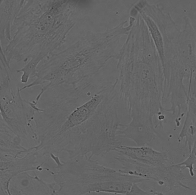
◆ Support for bright field imaging
Support real-time observation of cell microstructure in Mingchang
◆ Different magnification support
Supports 5X, 10X, 20X, 50X,100X and other magnification observations
◆ Electric load transfer
Electric two-dimensional loading platform, supporting live recording and observation of different culture dishes
◆ Electric focusing
Electric focusing, supporting multi position focusing recording
◆ Real time dynamic recording of cells
Recording real-time dynamics of living cells for long-term observation and recording of living cell processes
◆ Curve generation
Support various analysis curve generation functions
|
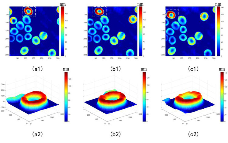
Real time dynamic recording of cells —— displacement of red blood cells at different times and 3D images |
||
|
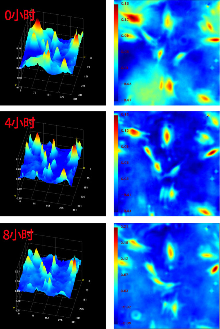
Real time dynamic recording of cells —— during the ESC live cell autophagy process Changes in cell state at 20x magnification: 0 hours, 4 hours, and 8 hours |
||
|
Cell 2D test results |
Cell contour map |
3D cell map |
No damage - no labeling or staining is required, no phototoxicity, and the true growth status of the recorded cells is restored.
Small size - can operate stably in the incubator for a long time and perform delayed imaging analysis under normal physiological conditions.
Real time monitoring - Real time monitoring of individual cells while obtaining population data and providing complete quantitative cellular morphological parameters.
Multiple data - can be reanalyzed for the same sample or result to obtain more experimental data.
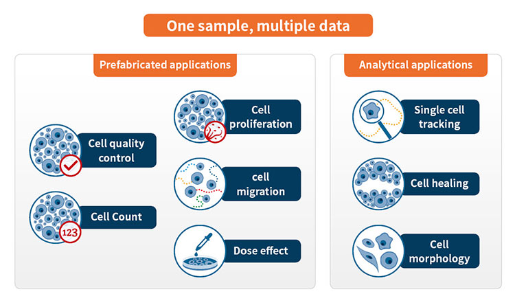
Widely applicable - can be used for research on cell proliferation, differentiation, migration, apoptosis, toxicology, and other aspects.
Intelligence - a concise and practical intelligent image analysis software that automatically performs high content quantitative analysis on the obtained images.
This BOJIONG Manual 3D Live Cell Imaging Microscope provides a fast, accurate, and non-invasive solution for automatic counting of adherent cells in incubators. Present images and results in real-time and clearly in a graphical manner, generate growth curves that change over time, and simultaneously monitor cell morphology, proliferation rate, and convergence.
Dynamic morphological observation of living cells
In the past, quantifying in vitro cell morphology was very difficult and time-consuming, as it involved inconsistent staining and the processing of complex cell imaging software. Unlike traditional cell imaging devices, BOJIONG Manual 3D Live Cell Imaging Microscope does not require phototoxic or faded fluorescent labels and can achieve non-invasive imaging of live cells and analyze various morphological characteristics, including individual cell volume, area, and thickness. Helps to visualize the differences between control cells and treatment cells, or to compare cell characteristics at different time points in the experiment.

BOJIONG Manual 3D Live Cell Imaging Microscope combines upright and inverted microscopes, combining the advantages of both. It is convenient and compact, with multiple functions in one machine. Users can easily switch between upright and inverted configurations through rotation, eliminating the need to purchase upright and inverted microscopes separately for different samples. One machine can image multiple samples, reducing costs and saving space.
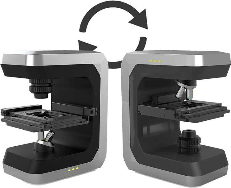
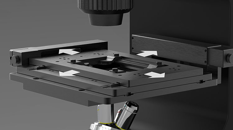
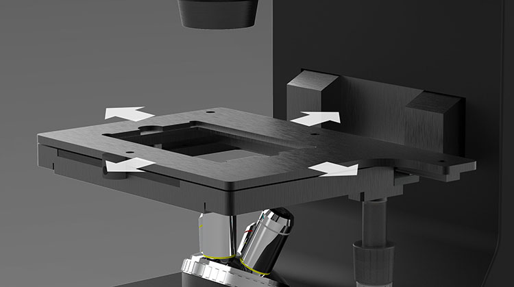
By flipping the main body of the microscope and flipping the fixed mobile platform, BOJIONG Manual 3D Live Cell Imaging Microscope can be flipped for easy magnification switching and observation in different occasions.

By flipping the main body of the microscope and flipping the fixed mobile platform, BOJIONG Manual 3D Live Cell Imaging Microscope can be flipped for easy magnification switching and observation in different occasions.
|
The mobile platform is fixed on the main body of the microscope through magnetic suction, and electric or manually controlled mobile platforms can be replaced as needed. |
|
|
|
Electric control stroke X:110mm,Y:75mm Table size:260mm×153mm |
|
|
|
Manual control stroke X: 190mm, Y: 90mm Table size:220mm×170mm |

This device can be equipped with microscope objectives of different magnifications: 5X, 10X, 20X, 50X, 100X; Observing the sample on an ultra-high definition display screen, it has unparalleled clarity.

BOJIONG Manual 3D Live Cell Imaging Microscope is equipped with powerful image analysis software, which is easy to use and includes precise image processing modules. It can not only provide population information of cells such as quantity, cell fusion degree, migration distance, etc., but also provide more than 30 morphological representations and motion parameters of individual cells, including area, thickness, volume, irregularity, etc., to extract quantitative data related to cells.
Address
No. 578 Yingkou Road, Yangpu District, Shanghai, China
Tel


")


")
